Thursday, 31 May 2012

Alcoholic extracts of Aloe vera for diabetic rats


This 2008 paper describes the antidiabetic activity of an Aloe vera gel extract with histological data to show the changes in STZ-induced diabetic rats.  This critique of the paper highlights the use of important data like extract doses (2004 J.Med. Food paper by Rajasekaran et al.), expected results, key hypotheses from earlier papers and the omission of important details such as the preparation of the different extracts of which the test extract was chosen as the best one.  I couldn't get the 2004 J.Med.Food paper but the 2006 paper in Clin. and Exper. Pharmacol. Physiol. by the same authors mentions some of the data in the 2004 paper. 
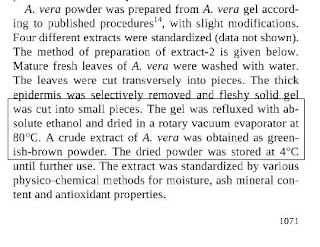
Four extracts made (DATA NOT SHOWN). Extract 2 taken for further study.  The gel is refluxed with absolute ethanol and dried in a rotary vacuum evaporator of 80 degrees C.  If alcohol can actually extract some of the polysaccharides in the gel, drying at 80 degrees C will cook this extract which is probably why the authors get greenish brown powder. 

 A 2008 review by C. T. Ramachandra and P. Srinivasa Rao in the American Journal of Agricultural and Biological Sciences (vol. 3, issue 2, pages 502-510) mentions that Aloe vera gel can be heated to 65 degrees Celsius for less than 15 min or to 85-95 degrees Celsius for 1-2 min.  Even if oxidation of certain compounds in the gel is important for antidiabetic activity, this heating at 80 degrees Celsius is a long one! 

Contrast this with the procedure described by Rajasekaran et al. in a 2006 paper.  The pulp (or gel) was lyophilized, extracted with 95% ethanol and dried (no temperature mentioned) to remove the solvent.  These authors also published a paper in 2004 in J. Med. Food which has been cited by Noor et al. in the 2008 paper.

------------------------------------------------------------------------------------------------------------
The 2006 paper by Rajasekaran et al. summarizes the reduction in blood glucose in STZ-induced rats receiving a dose of 300 mg/kg Aloe vera relative to control diabetic rats (21 days after administration).  The 2008 Noor et al. paper uses the same dose of 300 mg/kg Aloe vera extract (also three weeks) and gets approximately the same effect.  In fact, Rajasekaran et al. used 55 mg/kg bw STZ to induce diabetes, Noor et al. used 30 mg/kg STZ. 
                
------------------------------------------------------------------------------------------------------------
 Ghannam et al. (1986) used dried sap of Aloe vera and Rajasekaran et al. (2004, 2006) used 95% ethanol to extract Aloe vera pulp. 

-----------------------------------------------------------------------------------------------------------
Helal et al. (2003) already showed antidiabetic activity of Aloe vera in alloxan-treated diabetic rats. 

-----------------------------------------------------------------------------------------------------------
Would alcohol not extract the same principles water can PLUS other more non-polar molecules?  Could these kidney-related changes be due to the higher dose of STZ than due to alcohol used for the extraction?

This 2008 Noor et al. paper represents the increasing trend for peer reviewers to not have much time to read the manuscripts to do a thorough job and for the omission of important experimental details that will leave readers unable to reproduce much of the work described in the paper. 


Noor, A., Gunasekaran, S., Manickam, A.S. & Vijayalakshmi, M.A. (2008). Antidiabetic activity of Aloe vera and histology of organs in streptozotocin-induced diabetic rats, Current Science, 94 (8) 1078. DOI:

Sunday, 20 May 2012

Solution to ScFv to refolding

sciseekclaimtoken-4fc237fa76b9c
sciseekclaimtoken-4fbd15cda804b


<-----------------J. Biotech. 2011   (LEFT SIDE)                                                    

Sushma, K., Vijayalakshmi, M.A., Krishnan, V. & Satheeshkumar, P.K. (2011). Cloning, expression, purification and characterization of a single chain variable fragment specific to tumor necrosis factor alpha in Escherichia coli, Journal of Biotechnology, 156 (4) 244. DOI: 10.1016/j.jbiotec.2011.06.039


      J. Chrom B 2012 (RIGHT SIDE)      --------->                              

Sushma, K., Bilgimol, C.J., Vijayalakshmi, M.A. & Satheeshkumar, P.K. (2012). Recovery of active anti TNF-α ScFv through matrix-assisted refolding of bacterial inclusion bodies using CIM monolithic support, Journal of Chromatography B, 891-892 93. DOI: 10.1016/j.jchromb.2012.02.011


----------------------------------------------------

This section 3.2 is from the J. Biotech. paper and is an illustration of the rising trend of omitting experimental data which would be relevant to the study.                            


                                                                                        (J. Chrom B paper on the right)------>
 Should reviewers be OK with images of such quality?


J. Biotech paper (Figure 5)


----------------------------------------------------------------------------------------------------------
This paragraph is from the J. Biotech paper.  It appears that the ScFv from soluble fraction was less potent than Liu et al's anti-TNF alpha ScFv (already reported in 2007) and probably Geng et al's as well (?).

Why go for mutagenesis to bring something to a level that others have already shown or exceeded?  What are the advantages of E.coli over those expression systems used by Liu and Geng and their co-workers?

  ------------------------------------------------------------------------------------------------------------


Notice that in spite of the very close similarity of the assays and the data, the J. Biotech paper's Fig. 6 legend above states that "control (untreated in figure) cells were without any TNF-alpha and anti-TNF-alpha ScFv treatment."  Does this mean that control cells got anti-TNF-alpha treatment and not TNF-alpha?  

J. Chrom B's Fig. 4's legend states that "control cells were without any treatment."  As the results are so similar, would this mean that the above wording actually means this "without any treatment"? 

The results themselves show that untreated cells serve as the control for 100% viability in this MTT assay because they (as the 2012 J. Chrom B paper states clearly) are completely untreated. 

Why then are there points all along the X axis in the form of diamonds in the J. Biotech paper and in the form of crosses in the J. Chrom B paper? 

What concentrations are these and of what?
-------------------------------------------------------------------------------------------------------------


Duplicate (redundant) publication – refers to repeated publication of a manuscript when there is clear overlap with 
a previous manuscript already published. Such overlap is possible when there is distinct similarity in the basic assumptions underlying the research, the sample characterization, identity of patients, research methods, research findings or conclusions.


Salami publication- refers to splitting the research findings from a single study and dispersing them in different publications or at different times, with the intended purpose of increasing the number of publications. (Israel Medical Association)
-----------------------------------------------------------------------------------------------------------
The following two papers cover the expression, purification and characterization of a single chain variable fragment (ScFv) specific to TNF alpha in E. coli: one deals with the soluble fraction (J. Biotech, 2011), the other solubilizes the insoluble fraction (J. Chrom B, 2012). 
Analysis of both papers will be presented side-by-side (if space permits) or the figure of the (later) J. Chrom B paper will follow that of the (earlier) J. Biotech. paper to allow comparison. 

Would the J. Chrom B paper qualify as a salami or a redundant or just a me-too paper?
-----------------------------------------------------------------------------------------------------------


J. Biotech. 2011     (LEFT SIDE)                                                     J. Chrom B 2012 (RIGHT SIDE)

Sushma, K., Vijayalakshmi, M.A., Krishnan, V. & Satheeshkumar, P.K. (2011). Cloning, expression, purification and characterization of a single chain variable fragment specific to tumor necrosis factor alpha in Escherichia coli, Journal of Biotechnology, 156 (4) 244. DOI: 10.1016/j.jbiotec.2011.06.039


Sushma, K., Bilgimol, C.J., Vijayalakshmi, M.A. & Satheeshkumar, P.K. (2012). Recovery of active anti TNF-α ScFv through matrix-assisted refolding of bacterial inclusion bodies using CIM monolithic support, Journal of Chromatography B, 891-892 93. DOI: 10.1016/j.jchromb.2012.02.011


Saturday, 19 May 2012

Metal affects (enzyme) activity


What would the connection be between ROS in human cells and catalase from black gram as an enzymatic defense against ROS-mediated oxidative stress?  The authors imply that catalase in legumes can enter the gastrointestinal tract and get absorbed, intact, into the cells of the body to scavenge ROS?
The authors use Seph 4B-IDA-Zn(II) to purify the catalase.  Is there one band in lane 2 or are there bands very close to the middle band?  Could they have done HPLC or used some other method to show the purity of that component in lane 2?  Contrast to the paper by Ajila and Rao also given below. 

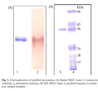
Ajila and Rao's paper:
Purification in this paper involves ion exchange chromatography on DEAE-Sephacel and gel filtration chromatography on Sephadex G100.

Their gels show a single band (real purification!) though the brightness is quite high. 
But they show the purity of their protein by doing HPLC and capillary electrophoresis.  Important in a chromatography journal to demonstrate the success of the chromatography done.

Back to Kandukuri et al, they test inhibition of catalase with different metals and inhibitors, much like Ajila and Rao (Table 6 below).  However, in spite of using Zn(II) for their IMAC, Kandukuri omit to test the effects of Zn(II) on catalase activity.  Enzyme inhibition is not the only aspect of interest, induction of enzyme activity is also an important aspect to consider. 
According to Ajila and Rao, ZnCl2 treatment actually gave 120% activity compared to a (normalised) 100% of control (untreated). 


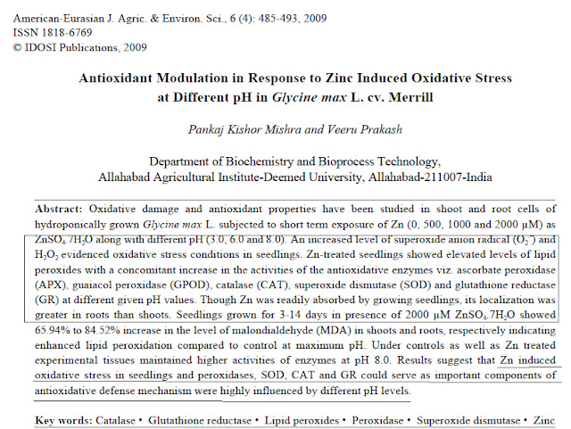
 Another paper to look at is by Mishra and Prakash (given below as abstract):                         
Mishra and Prakash's paper:
Here too, it is shown that Zn-treatment causes accumulation of the metal in the roots accompanied by increased oxidative stress due to which antioxidant enzymes like peroxidases, SOD, catalase, GR etc are induced. 

Bottomline: On the one hand, you have reviewers who hardly spend any time understanding any experiments in the manuscript and casually propose experiments that are either out of scope or are too much of an investment compared to the return that publication in that journal would give. 
Here, however, some questions have not been asked by the reviewers. 


Kandukuri, S.S., Noor, A., Ranjini, S.S. & Vijayalakshmi, M.A. (2012). Purification and characterization of catalase from sprouted black gram (Vigna mungo) seeds, Journal of Chromatography B, 889-890 54. DOI: 10.1016/j.jchromb.2012.01.029


Ajila, C., & Prasada Rao, U. (2009). Purification and characterization of black gram (Vigna mungo) husk peroxidase Journal of Molecular Catalysis B: Enzymatic, 60 (1-2), 36-44 DOI: